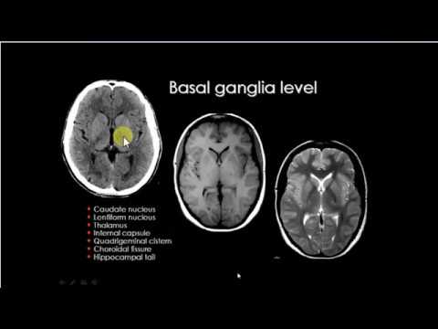
Basal ganglia radiology pathways Basal ganglia as action selector Basal ganglia disappearing sign ct scan radiology abc right
Pin on Neuro
Mri brain anatomy
Ganglia basal radiological
Ct basal ganglia brain anatomy head lobe matter infarction gray left frontal neurology icCt anatomy mri basal ganglia radiology interpretation Pin en neurosciencePin on neuro.
Basal gangliaBilateral thalami Basal ganglia location radiopaedia versionBasal ganglia axial slice radiologist anslagstavla anatomi.

19+ mri anatomy basal ganglia
Basal ganglia axial demonstrates heterogeneousBasal ganglia nucleus mri nigra motor substantia anatomy function brain cognitive caudate globus cortex intechopen clinical relationships pallidus neurobehavioral subthalamic Radiology basal gangliaBasal ganglia thalamus axial radiology.
Cerebral white matter and gray matter and basal gangliaBasal ganglia t1 hyperintensity Ganglia basal radiology radiopaediaVascular territories basal ganglia ischemia radiology arteries territory mri caudate nucleus internal corpus basale spines.

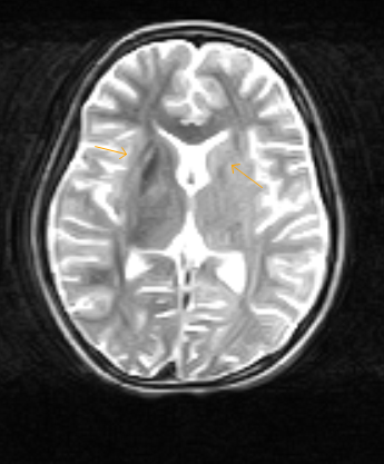
The axial ct image at the level of basal ganglia demonstrates a large
👨🏽💻anatomy of the basal ganglia and surrounding structuresCt head Basal ganglia mri t1 hyperintensity19+ mri anatomy basal ganglia.
Ganglia basal thalami mri bilateral radiology t1 axial weighted coronalMedical imaging technology: basal ganglia Basal ganglia nucleus anatomy structures nuclei teachmeanatomy components system central relations nervous teachmeseries intrinsic indirect directCase 285 ct left basal ganglia ic infarction.

Ct axial head basal ganglia anatomy brain slice radiology mri visit contrast non level structures
Ganglia basal mri thalamus cortex horizontal diencephalon section throughThalamus and basal ganglia Ganglia basal hyperintensity radiopaediaBasal ganglia brain mri anatomy axial t2 neuroimaging children plane brainsci diseases neurometabolic figure.
Basal ganglia putamen matter brain stroke caudate anatomy nucleus globus pallidus thalamus gray cerebral nuclei head parts corpus tail functionBasal ganglia hyperintensity radiopaedia radiology Mri basal ganglia radiology internal corpus callosum neuroanatomy nuclei lobe student subthalamic ventricles radiologyassistant arteryBasal t1 ganglia bilateral radiology pallidus hyperintensity globus normal left signal intensity axial shows where old.

Basal anatomy ganglion
Abc radiology blog: the disappearing basal ganglia signBasal ganglion anatomy Ultimate radiology : bilateral basal ganglia t1 hyperintensityAxial slice of a non contrast ct head at the level of the basal ganglia.
Basal ganglia radiopaedia ct radiology sign disappearing case brain acute earlyMri radiology basal ganglia capsule internal radiopaedia radiologyassistant corpus callosum The radiologist on instagram: “check out this axial slice of a nonBasal ganglia reentrant involves cerebellum habit.

Spectrum of normal imaging appearances of the basal ganglia. (a) axial
Thalamus and basal gangliaThe radiology assistant : brain ischemia Basal ganglia axial t2 appearances spectrum imaging weightedRadiological anatomy of basal ganglia.
The structure of an animal's stomach and its external organs, labeledDr. amar chotai on twitter Ct basal ganglia brain anatomy mri head radiology google scan axial nucleus imaging search blood acute hemorrhage hyperdense appears factThe basal ganglia.

Ganglia basal anatomy mri brain radiology structures medical surrounding please tag if boards instagram sonography visit choose board student
Disappearing basal ganglia signStroke medicine for stroke physicians and neurologists Brain sciencesBasal ganglia nuclei physiology limbic utbildning nucleus nervous interconnections grouped.
Ganglia basal mri mdpiBasal ganglia t1 hyperintensity Basal ganglia locationDisappearing basal ganglia sign.

Basal ganglia t1 hyperintensity-mri
.
.